Product Description
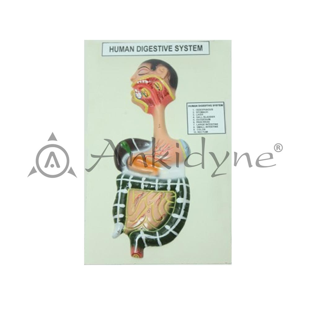
The Human Digestive System Model is a highly detailed, colourful, three-dimensional relief teaching aid mounted on a cream/off-white display board, designed to provide a complete and accurate visual representation of the entire human digestive tract from the oral cavity to the rectum. Titled "HUMAN DIGESTIVE SYSTEM" at the top, this wall-mount/tabletop model is an essential classroom and laboratory tool for teaching biology students about the structure, sequence, and function of all digestive organs.
The model features a realistic cross-sectional head profile showing the oral cavity, teeth, tongue, salivary glands, pharynx, and oesophagus in vivid detail, seamlessly transitioning into the abdominal digestive organs including the liver (dark green), stomach, gallbladder, duodenum, pancreas, small intestine, large intestine, colon, and rectum all rendered in anatomically accurate colours and positions. A labelled identification chart titled "HUMAN DIGESTIVE SYSTEM" listing 10 numbered structures is affixed to the right side of the board for easy classroom reference.
Realistic, Educational 3D DesignThis wall-mountable model features lifelike anatomical details and multi-colored, glossy painted organs, bringing the human digestive system to life. Its relief format provides tactile, three-dimensional perspectives, making anatomy lessons interactive and visually engaging for students.
Durable and Low-MaintenanceManufactured with strong plastic/PVC, the model is built to withstand frequent handling. Its surface resists cracking and cleaning is simple, which ensures hygienic use in classroom settings. The included box protects the product during transportation and storage, extending its service life.
Versatile Teaching ToolDesigned for use in school laboratories, study rooms, and biology lectures, this wall-mounted digestive system model supports various teaching methods. It helps students visualize organ positions and relationships, fostering better understanding through demonstrations and collaborative study.
FAQ's of Human Digestive System Model:
Q: How is the Human Digestive System Model used in educational settings?
A: Teachers and learners utilize the wall-mounted model during biology classes and laboratory sessions to visually demonstrate the structure and functions of the human digestive system, making complex anatomy concepts easier to understand.
Q: What anatomical features are displayed on this model?
A: The model accurately represents the mouth, oesophagus, stomach, small intestine, large intestine, rectum, liver, pancreas, and gall bladder, showcasing their shapes, positions, and relationships in the digestive tract.
Q: When is this model most beneficial in the learning process?
A: It is especially effective during anatomy lessons, practical demonstrations, and examination preparations, enhancing students' comprehension of digestive system organization and physiology.
Q: Where can the model be installed for optimal learning impact?
A: Ideal installation locations include biology classrooms, school laboratories, and study rooms. Its wall-mountable design ensures visibility and accessibility for group study or lectures.
Q: What is the process for cleaning and maintaining the model?
A: The model's plastic/PVC construction and glossy finish make maintenance easy. Simply wipe it with a damp cloth to remove dust or marks, ensuring its longevity and hygiene in shared educational spaces.
Q: What are the key benefits of using this anatomical model?
A: It offers hands-on, visual interaction with digestive system anatomy, fosters deeper engagement, supports multiple teaching methods, and withstands regular classroom use due to its durable materials.
Q: How is the model packaged and protected during shipment?
A: Each model is securely box packed to prevent damage, ensuring it arrives safely for immediate classroom or laboratory use.